We build foundation models that power imaging devices across science, medicine and industry.
Pushing sensors to the limit.
Same hardware, faster imaging.
Quantitative physics-aware models.
3D-native reconstruction.
Instantly generalise to new deployments.
Seamless in existing pipelines.
Built for the most regulated industries.
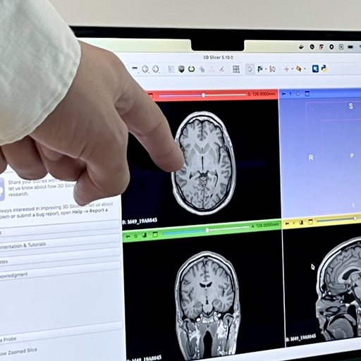
As MRI accelerates and develops, clinical acquisitions face myriad artifacts: aliasing caused by undersampling spokes or lines, B0 inhomogeneity sensitivity and off-resonance due to long readouts, low SNR or resolution at ultra low-field, or temporal bias and motion artifacts in free-breathing sequences. Our models operate in a new paradigm of MRI: one product to reconstruct anything, in any country, grounded in any acquisition physics.
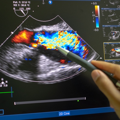
Today's ultrasound pipelines rely on hand-crafted beamforming and post-processing, discarding precious RF data. They hence limit resolution and contrast and struggle with complex tissue propagation, speckle or aggressive subsampling. Our foundation models solve the full ultrasound inverse problem that enable new forms of real-time, fully 3D, quantitative ultrasound exams.
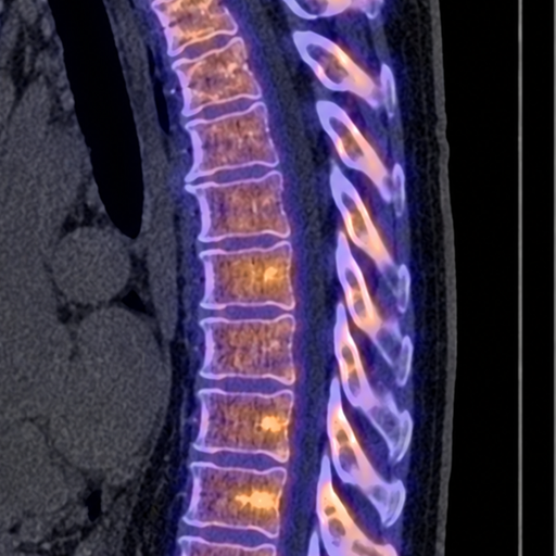
Clinical SPECT acquisition is characterised by challenging noise statistics, PVEs, collimator blur, and angular subsampling, and today's classical reconstruction methods cause noisy images, reduced lesion detectability, biased quantification, and low diagnostic confidence. The next generation of SPECT low-dose and accelerated imaging for theranostics forces clinicians to trade acquisition time and dose with image quality. Our models are being used to reconstruct images of unprecedented quality and quantitative consistency.
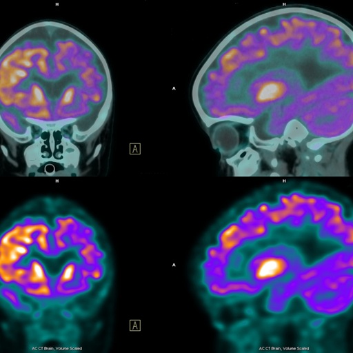
In PET, huge, expensive hardware or long acquisition times are traditionally needed to collect enough photons and reconstruct images. Even so, lesion detectability, quantification and diagnosis still suffer due to weak reconstruction algorithms, poor ToF modelling or challenging detector geometries. Our models reconstruct images of unprecedented quality while preserving SUV consistency, enabling next-order throughput cancer imaging at a tiny fraction of the cost.
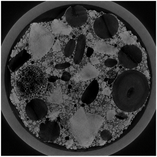
Non-destructive testing requires large 3D volumes with high resolution and fine texture fidelity to detect defects. However, acquisition and reconstruction times are major bottlenecks in production environments. Accelerated sparse-view or limited-angle protocols introduce streaking, reduced contrast, and loss of fine structural detail. Our models solve CT image reconstruction from any geometry of incomplete or noisy radiographs, enabling faster inspection of critical details in images.
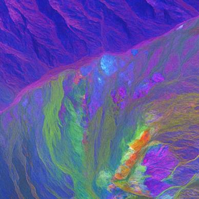
In any optical image sensor, raw measurements are shaped by spectral sensitivity, CFAs, exposure, optics, and noise, and the sensor cost trades with image resolution and SNR. This trade-off is bottlenecked by traditional ISP pipelines, where classical deblurring or demosaicing methods blocks discard precious information and downgrade the spatial-spectral resolution. Our foundation models reconstruct machine-vision data directly from raw sensor measurements, enabling new forms of high spatial-spectral resolution imaging with cheaper, smaller cameras, without needing any large paired training datasets.
Fluorescence microscopy is limited by the trade-off between resolution, speed, field of view, phototoxicity, and photon budget. Low-light or fast acquisitions reduce bleaching and enable live imaging, but produce noisy, blurred, or incomplete measurements. Our models have been demonstrated by microscopists to restore images from degraded data, preserving biological structure and quantitative fidelity, so that microscopists can spend time analysing images, not correcting for poor image quality.
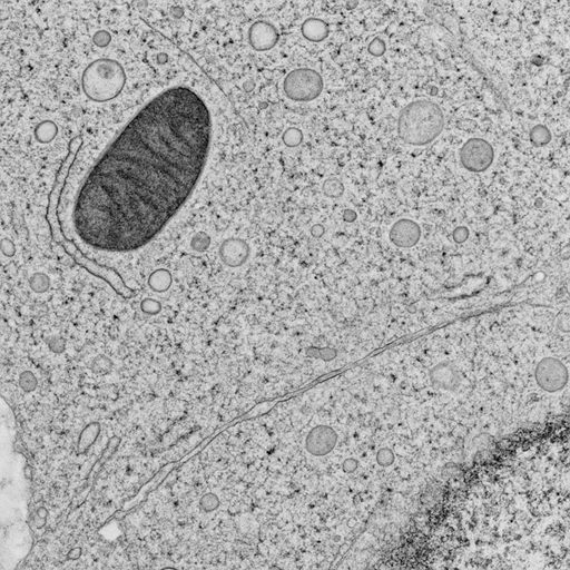
Electron microscopy and tomography trade dose, dwell time, resolution, and field of view against beam damage and detector physics. Low-dose or accelerated acquisitions leave measurements degraded by shot noise, scan distortion, charging, drift, beam-induced motion, frame misalignment, and angular undersampling. Our foundation models reconstruct micrographs and tomograms from dose-limited, distorted, or incomplete measurements, accounting for acquisition timing and specimen motion, recovering structural fidelity across any electron imaging modality.

RAM presented at ICLR 2026
Read more ›
2 papers presented at CVPR 2026
Read more ›
Meet Blur Labs at our VivaTech stall
Read more ›
The final frontier of imaging at PyTorch Conference Europe
Read more ›
Press published by University of Cambridge Engineering News
Read more ›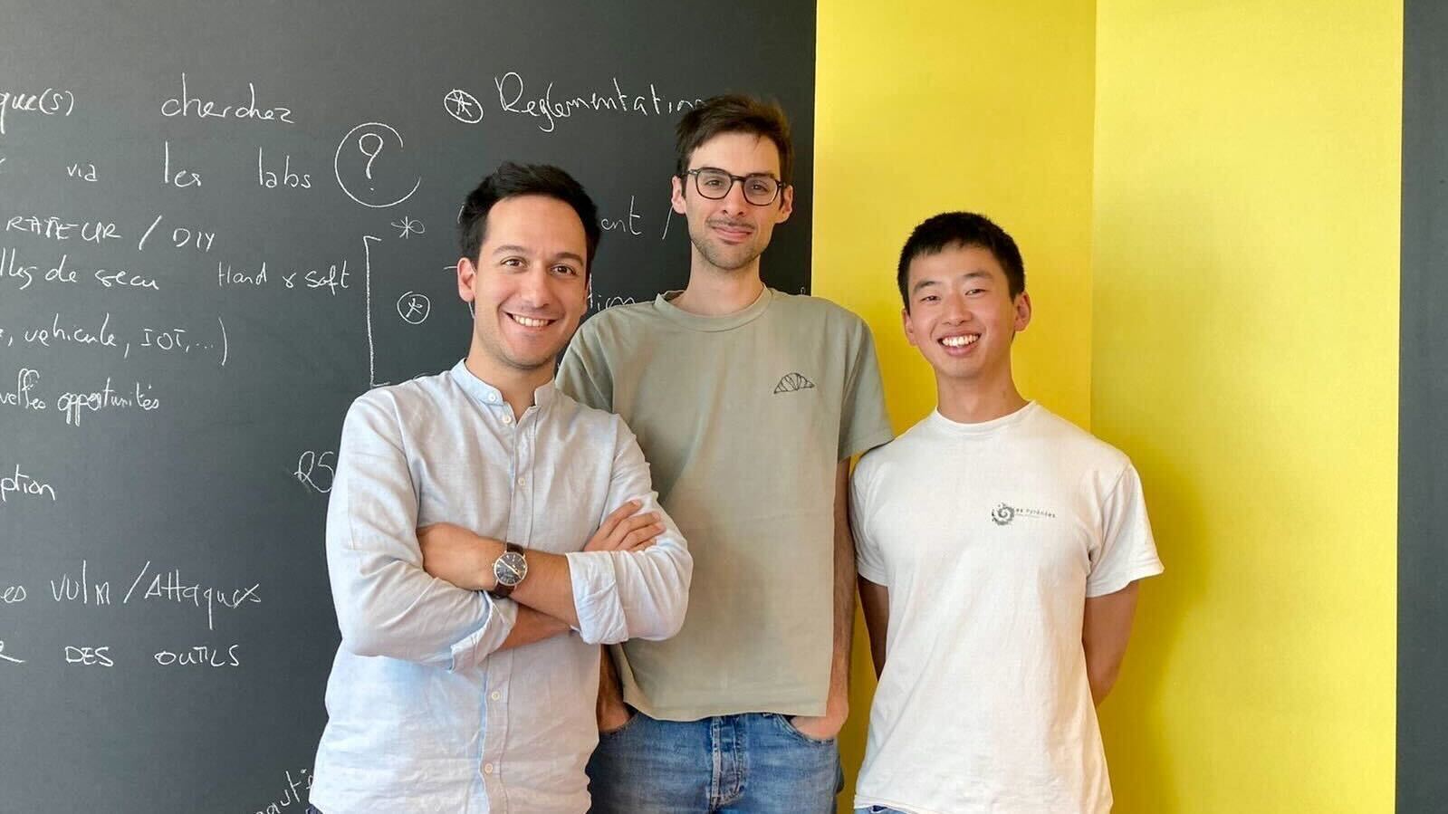
Founding team joins Inria Startup Studio
Read more ›